Advanced Medical Imaging: Transforming Cancer Diagnosis and Treatment
In today’s fast-evolving medical landscape, the role of advanced medical imaging has become super important in supporting every stage of cancer care. The ability to see inside the body, from the early detection of cancer to monitoring treatment outcomes, has changed the way physicians work and how patients experience their journey. Medical imaging now spans a range of techniques, from digital breast imaging to nuclear medicine scans—all designed to offer precise, detailed insights while helping patients and care teams steer through the often overwhelming treatment process.
This opinion piece explores various radiology techniques, highlighting their benefits, how they have evolved, and what patients can expect from them. We’ll take a closer look at digital breast imaging, computed tomography, interventional radiology, and more. Each imaging modality offers a unique perspective into the fine points of cancer care and treatment—a factor that is both critical and must-have for ensuring the right interventions at the right time.
Innovative Imaging in Cancer Care: Digital Breast Imaging and Beyond
When it comes to addressing breast cancer, the methods available today have evolved dramatically. Digital mammography, often using 3D mammography (or digital breast tomosynthesis), has literally revolutionized how early anomalies are spotted. With the ability to take multiple images layer by layer, radiologists are better equipped to identify slight differences in tissue density that may signal early changes.
A breast MRI complements this approach by creating detailed images using strong magnetic fields rather than X-rays. The advantage here is that the procedure can reveal small, subtle parts of the breast tissue that might be hidden during a conventional exam. In some situations, a breast ultrasound also comes into play. Ultrasound relies on sound waves to craft pictures of tissue structures, and is particularly useful when determining whether a mass is solid or fluid-filled. These methods together offer a wholesome safety net, ensuring that both patients and their providers have multiple angles from which to evaluate potential concerns.
The use of contrast agents—whether injected or consumed—further enhances imaging quality. Contrast helps highlight specific tissues and can be the key to visualizing the tricky parts of tissue changes that standard images might miss. With these techniques, doctors can get into the nitty-gritty of breast cancer detection, ensuring that even the tiniest deviations are not overlooked.
Some advantages of advanced breast imaging include:
- Enhanced image detail: 3D mammography provides clear, layered images to reveal small differences in tissue.
- Multimodal approach: The integration of ultrasound, MRI, and digital imaging allows for a thorough examination of anomalies.
- Improved diagnostic accuracy: Contrast-enhanced imaging deepens the clarity of specific tissues, unlocking the fine details that are essential for early detection.
These advancements not only offer improved diagnostic capabilities but also reduce the nerve-racking uncertainty that patients might feel during the screening process.
Tackling the Tricky Parts: Understanding CT Scans and Interventional Radiology
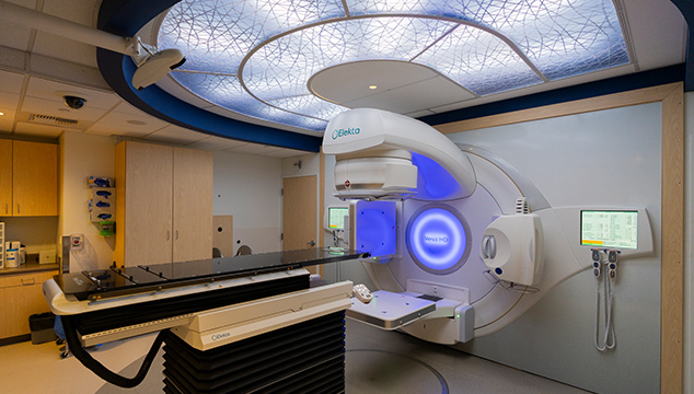
Computed tomography (CT) scans are another cornerstone in cancer care, providing detailed three-dimensional images of the inside of the body. These scans allow doctors to see organs, bones, and blood vessels with precision, making it easier to plan specific treatments. For example, when patients are gearing up for radiation therapy, CT simulation scans are usually the first step. These crucial steps help the care team figure a path to pinpoint the area needing treatment.
During a CT scan, patients lie on a table while an X-ray tube animates around them, capturing cross-sectional views of the body. Often, a contrast agent is used to shed light on soft tissues, enhancing the quality of the images. This approach is particularly useful for diagnosing cancers and determining the stage of progression, ensuring that the extent of the disease is as transparent as possible.
Interventional radiology represents another innovative branch of imaging. Physicians trained in these techniques use real-time imaging guidance to perform minimally invasive procedures. Whether it’s a needle biopsy to extract a tissue sample or thermal ablation to destroy tumor cells, interventional radiology offers less invasive alternatives to traditional surgery. By using imaging technologies such as ultrasound, CT, or X-ray fluoroscopy, doctors can carefully direct instruments to the precise target area without the need for extensive incisions.
Key benefits of CT scans and interventional radiology include:
- Accurate disease staging: CT scans provide multidimensional images that clearly show the extent of cancer, guiding further treatment.
- Minimally invasive procedures: Interventional radiology techniques reduce recovery times and minimize complications.
- Real-time guidance: With live imaging, physicians can easily adjust treatments in the moment, ensuring precision.
By addressing the confusing bits of traditional diagnostic methods, these imaging solutions allow both physicians and patients to breathe easier, knowing that every twist and turn of the disease process is being carefully monitored.
Getting Into the Details: MRI and the Debate Over Whole-Body Scanning
Magnetic resonance imaging (MRI) has long been a trusted ally in the diagnostic process, especially when it comes to visualizing soft tissues. Unlike CT scans that rely on X-rays, MRI uses a combination of a strong magnet and radio waves to capture highly detailed, three-dimensional images. This method is particularly valuable for detecting tumors, assessing their spread, and planning subsequent treatments.
An interesting debate in the field centers around whole-body MRI scans. While some proponents tout these as a way to screen for cancer in those currently showing no symptoms, the current consensus among experts is that whole-body MRI is not a recommended screening tool for the general public. The main concern is that these scans may uncover harmless anomalies—false alarms that could lead to additional, and sometimes intimidating, follow-up procedures.
The fine points involved in choosing the right imaging method can be challenging. While whole-body MRI can potentially pick up minute details, the risk of generating unnecessary anxiety and triggering further testing means that it is not widely endorsed as a routine screening tool. Instead, targeted MRIs that focus on areas of concern are seen as super important for ensuring that patients receive the right level of attention and care.
A closer look at magnetic resonance imaging reveals several notable benefits:
- High-resolution images: MRI is excellent at revealing soft tissues and small distinctions within the same organ.
- Non-invasive evaluation: Without the use of harmful radiation, MRI provides a safe way to examine subtle tissue changes.
- Customizable protocols: MRI testing can be tailored to the specific needs of the patient and the suspected condition.
Understanding these techniques helps demystify what might appear as intimidating technology, making it easier for patients to get around the complicated pieces of their treatment journey.
Modern Nuclear Medicine: Tracing Functions Beyond Structures
Nuclear medicine is one of those areas where imaging does more than simply map out the body’s blocks and tissues. Instead, nuclear medicine scans like PET/CT and PET/MRI focus on both structure and function. By administering a small amount of a radioactive substance—known as a radiotracer—physicians can track how cancer cells absorb the tracer differently from normal cells. This process provides a dual-layer insight: not just showing the physical layout of organs, but also the functional performance of tissues.
A PET/CT scan is particularly noteworthy due to its combined approach. The CT component of the scan provides the detailed structural image, while the PET portion reveals metabolic activity within the tissues. This merged view enables a more comprehensive understanding of cancer behavior. In some cases, SPECT/CT scans are used, especially in scenarios where patients are receiving molecular therapies such as targeted drug delivery that employs radiation directly to the tumor.
Nuclear medicine offers several advantages:
- Dual diagnostic capabilities: The combination of anatomical and functional data makes images more informative.
- Enhanced detection of disease: These scans can reveal subtle metabolic changes that might not be visible on standard imaging.
- Tailored treatment approaches: Insights from nuclear imaging help physicians design personalized treatment plans that address both the physical and functional aspects of cancer.
By providing that extra layer of detail, nuclear medicine not only increases diagnostic accuracy but also helps in planning further interventions with a clearer understanding of the hidden complexities behind tumor behavior.
Ultrasound: A Non-Invasive Guide in Diagnosis and Treatment
Ultrasound imaging, though one of the most traditional techniques in the medical toolkit, remains a critical component of modern diagnostic procedures. This method employs sound waves to create images of the internal structures of the body. It is particularly useful because it exposes patients to no radiation, offering a safe and non-invasive way to assess tissues and organ function.
In the context of cancer care, ultrasound is versatile. It can help determine if suspicious masses are solid or fluid-filled, guide needle placements during biopsies, and even assist in placing markers for precise radiation targeting. The technology is especially valued for its real-time imaging capabilities, allowing care teams to see the effects of an intervention as it happens.
Here are some of the key uses of ultrasound in cancer diagnostics:
- Real-time imaging: Ultrasound provides instant feedback during procedures, which is critical for accurate needle placements and interventions.
- Versatile application: From detecting fluid collections to assisting with biopsies, ultrasound fills several diagnostic gaps.
- Non-invasive approach: As it does not involve radiation, ultrasound is a safer alternative for repeated examinations.
By incorporating ultrasound into imaging routines, healthcare providers are better able to sort out the subtle differences in tissue structure without exposing patients to additional risks. This blend of safety and efficiency makes ultrasound a cornerstone of modern cancer diagnostics.
Handling the Overwhelming Process: What to Expect During Radiological Examinations
For many patients, the process of undergoing radiological exams can feel intimidating. This is especially true if you are facing a cancer diagnosis, where every moment seems loaded with emotion and uncertainty. However, understanding the process and what to expect can help ease those overwhelming concerns.
Before your imaging appointment, your healthcare team will explain why a specific scan is recommended and what the procedure will entail. Whether you are scheduled for a CT, MRI, or ultrasound, it’s important to remember that these procedures are designed to provide a clear picture of your internal structures. Radiologists and technicians are trained to guide you through every step—from preparing for the scan (which might include fasting or avoiding certain medications) to understanding the images that are generated.
Patients are often advised to:
- Ask questions: Don’t hesitate to inquire about the procedure’s details, including how you should prepare and what sensations you might experience.
- Follow pre-scan instructions: Specific instructions might include wearing comfortable clothing or arriving with an empty stomach if contrast material is used.
- Relax and communicate: If you are feeling nervous, let the staff know. They can help alleviate your concerns and offer modifications to ensure your comfort.
By taking the time to explain each step and establish clear communication channels, radiology departments help patients figure a path through the intimidating maze of medical imaging. This collaborative approach fosters confidence and builds trust with your care team, ensuring that you feel informed and supported.
Maintaining Focus: Safety and Quality in Radiological Practices
In the realm of advanced medical imaging, safety and quality are not just buzzwords—they are essential pieces of each diagnostic process. Institutions dedicated to cancer care have implemented rigorous protocols to ensure that while the imaging techniques are cutting-edge, they also adhere to the highest safety standards. The right balance between accurate, detailed imaging and minimal risk exposure is key.
Quality assurance in radiology involves multiple checkpoints. From the calibration of imaging machines to the ongoing training of the radiology staff, every element is designed to reduce errors and maximize the clarity of the images produced. Some of the critical safety protocols include:
- Regular equipment maintenance: Advanced machines are checked and recalibrated frequently to guarantee optimal performance.
- Technician training: Continuous education ensures that staff are up to date on the latest imaging techniques and safety procedures.
- Patient-specific protocols: Tailoring procedures to individual patient needs helps lower risks, especially when contrast agents or radiation are involved.
These procedures not only enhance the reliability of the imaging tests but also help to mitigate the potential twists and turns that can come with complicated pieces of treatment. Knowing that the imaging process is both safe and of high quality can offer significant peace of mind to patients who are already juggling the scary aspects of their diagnosis.
Radiology in the Broader Spectrum of Cancer Rehabilitation and Patient Care
While advanced imaging techniques play a vital role in diagnosis and treatment planning, they also contribute significantly to the overall approach to cancer rehabilitation and patient care. Radiology is intricately tied to other supportive services in a comprehensive cancer care program. It doesn’t operate in isolation; instead, it helps guide a broader strategy that includes integrative medicine, nutritional planning, physical therapy, and mental health support.
For many patients, the journey doesn’t stop at diagnosis. Post-treatment monitoring and follow-up imaging are super important to assess how well the body is healing and to spot any signs of recurrence early. This ongoing relationship between imaging and patient care helps ensure that when it comes time to sort out the subtle, confusing bits of recovery, the right tools and strategies are already in place.
Here’s how radiology is integrated into broader patient care:
- Monitoring treatment effectiveness: Regular scans help physicians see if a treatment is working or if adjustments are needed.
- Supporting rehabilitation: Imaging can guide physical therapy and other recovery programs by showing progress or new areas that may need additional care.
- Guiding follow-up care: Continued imaging ensures that any new concerns are detected promptly—an essential part of long-term survivorship strategies.
This integrated approach exemplifies how imaging is one part of a multi-channel care system that considers every layer of a patient’s well-being. It not only assists in deciphering the physical changes but also supports the management of emotional stress and the nerve-racking uncertainties of cancer recovery.
Looking Ahead: The Future of Radiology in Cancer Care
The future of radiology in cancer care is filled with promise and innovation. Researchers and technology developers are continually refining imaging techniques, developing new contrast agents, and even exploring artificial intelligence to assist in image reconstruction and interpretation. This means that in the near future, diagnoses may occur even faster, treatments may be tailored more precisely, and the overall patient experience might be transformed for the better.
One trending area is the integration of AI algorithms that can analyze images for subtle differences that might welcome further investigation. These algorithms are designed to help professionals sift through the little details that are sometimes missed by the human eye. Additionally, as more data becomes available, imaging protocols will be better able to adjust to individual patient characteristics—further personalizing the approach to cancer care.
What does this mean for patients and healthcare providers? For one, improved diagnostic precision leads to more individualized treatment plans, which in turn, can improve outcomes and reduce unnecessary procedures. It also helps in easing the anxieties that come with uncertainty, as patients can be reassured that every possible twist and turn is being thoroughly examined.
In summary, the future of radiology promises:
- Advanced image analysis: AI and machine learning will enhance the ability to spot subtle indicators of disease.
- Improved personalization: Tailored imaging protocols based on individual health profiles will lead to better patient outcomes.
- Streamlined patient care: Faster and more accurate imaging will help reduce the waiting times and nerve-racking delays in treatments.
As these advances continue, the fine points of radiology will only become more critical in supporting the health and well-being of cancer patients, ensuring that every stage of the care process is underpinned by the latest technological innovations.
Conclusion: Steering Through the Future of Cancer Care with Confidence
The evolving landscape of radiology is continually proving to be a cornerstone in modern cancer care. From digital breast imaging and CT scans to interventional procedures and nuclear medicine, these advanced techniques offer patients and care teams the tools needed to get around the confusing bits and tangled issues of cancer diagnosis and treatment. By providing detailed images and functional insights, radiology helps untangle the complexities that have traditionally made cancer care a nerve-racking experience.
Moreover, the collaborative approach between imaging specialists and the broader care team ensures that every step—whether it involves guiding a needle for a biopsy or monitoring how well a treatment is working—is done with the utmost precision and care. Patients can feel confident knowing that behind each scan lies a commitment to high safety standards, continuous technological innovation, and an unwavering focus on improving outcomes.
As we move further into an era of personalized medicine, the role of advanced imaging becomes even more central. With every innovation, from AI-assisted analysis to improved contrast imaging, those charged with diagnosing and treating cancer are better equipped to figure a path through the twists and turns of the disease. This means not only a clearer view of what lies inside but also a more supportive, patient-centered approach to care.
Ultimately, advanced medical imaging is more than just a diagnostic tool—it is a key partner in guiding patients through every stage of cancer care. By combining cutting-edge technology with thoughtful, patient-centered procedures, radiology continues to pave the way for a future where early detection, precise treatment guidance, and effective follow-up care converge to offer hope, clarity, and improved health outcomes.
For patients and providers alike, the message is clear: while the journey through cancer care may be loaded with issues and subtle details that require careful interpretation, advanced imaging is here to help make the complicated pieces a bit clearer. With an ever-expanding suite of diagnostic tools, radiology stands as an essential force driving both innovation in treatment and the betterment of patient quality of life.
In a world where timely, accurate diagnoses are super important, it is heartening to see that medical imaging is stepping up to the challenge. By merging technology with compassionate care, radiology not only addresses today’s pressing needs but also paves the way for tomorrow’s breakthroughs in healthcare. Through continual research, refinement, and an unwavering focus on patient well-being, the field is poised to ease the intimidating aspects of cancer treatment while unlocking new possibilities in medicine.
As we look to the future, it is crucial for healthcare providers, researchers, and policymakers to continue investing in state-of-the-art imaging technologies. Doing so will ensure that our health systems remain capable of tackling the ever-evolving, tricky parts of cancer diagnosis and treatment. With every scan, every image, and every analyzed detail, we are one step closer to a world where cancer is detected earlier, treated more effectively, and managed in a way that reduces the overall load of uncertainty for patients.
In conclusion, advanced radiology is indispensable in modern cancer care. It is not only about capturing images but also about guiding patients and doctors through every complicated piece of the treatment process. As the technology matures and becomes more integrated into the fabric of healthcare, the day when every patient can feel fully supported through their diagnostic journey grows ever nearer. By embracing these innovations, we are not just creating images but building hope—one scan at a time.
Originally Post From https://www.fredhutch.org/en/patient-care/treatments/radiology.html
Read more about this topic at
Innovative Imaging Techniques for Advancing Cancer …
Advanced Cancer Imaging Programme