![]()
![]()
An Opinion on the Future of Cancer Imaging
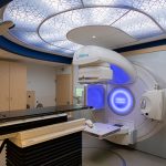
Advances in radiology have changed the way we figure a path through cancer care. As someone who has spent many years observing modern and alternative methods of healing and treatment, I find the role of sophisticated imaging both inspiring and essential. Radiology techniques, including those offered at institutions like Fred Hutch Cancer Center, provide patients and health care professionals with tools to dig into the fine points of cancer treatment. Today, I want to share my views on how these imaging methods, although sometimes tangled with tricky parts and nerve-racking complexities, are transforming the entire landscape of oncology care.
Over the years, we have watched traditional diagnostic methods blend with cutting-edge imaging technologies. This blend creates a more comprehensive overview that helps doctors and patients alike make informed decisions. I plan to take a closer look at several imaging modalities and explore their role in screening, diagnosis, treatment planning, and follow-up of cancer care.
Understanding Radiology Techniques in Cancer Care
Radiology is more than just a set of tests; it is a vital window into the inner workings of our bodies. The imaging methods used in cancer treatment allow the health care team to see the tricky parts hidden inside the body, revealing subtle details and little twists that might be missed in a traditional physical exam.
Let’s dive in and explore some common imaging techniques, how they work, and why they are so popular at advanced centers like Fred Hutch.
Breast Imaging: Modern Approaches for Early Detection
Early detection is key when figuring a path to better health, and breast imaging stands as one of the most super important tools in this battle. At centers such as Fred Hutch, the use of 3D mammography, or digital breast tomosynthesis, means that images are captured layer by layer, enhancing the view of the breast tissue in a way that traditional techniques cannot match.
This process involves using X-rays to create detailed shots of the breast. The technology is built to enhance the subtle parts of an abnormal tissue by superimposing several layers of images. Occasionally, the imaging process may require a contrast agent – a substance that is introduced into the body to reveal otherwise hidden details. In some cases, a breast magnetic resonance imaging (MRI) scan is used, offering another angle to get into the nitty-gritty of breast tissues, particularly in complex cases.
- 3D Mammography: Offers a layered view of tissues for better early detection.
- Breast MRI: Uses magnetic fields instead of X-rays and is ideal for patients with dense breast tissue.
- Breast Ultrasound: Provides additional insights, especially when trying to figure out if a mass is solid or filled with fluid.
The combination of these tools not only boosts the accuracy of cancer detection but also reduces the likelihood of unnecessary invasive procedures. This integrated approach is absolutely key in fighting the overwhelming challenge of cancer.
Computed Tomography (CT) Scan: A Closer Look at Internal Structures
The CT scan is another cornerstone of modern diagnostic imaging. Using a special X-ray machine, CT scans take detailed cross-sectional images that portray both organs and bones with fine shades of detail. For anyone dealing with cancer, seeing organs clearly can be the difference between early detection and a delayed diagnosis.
Before the procedure, a contrast material may be administered to spotlight blood vessels and tissues. This helps doctors figure out a detailed map of the area, making it easier to design personalized treatment plans. In fact, many cancer patients undergo CT scans not only for initial assessment but also for planning subsequent treatments like radiation therapy. When it comes to certain cancers, low-dose CT scans have become a super important screening tool, particularly for those at high risk of lung cancer.
Magnetic Resonance Imaging (MRI): The Magic of Magnets
MRI offers a different approach by using powerful magnets and radio waves to capture detailed 3D images. Unlike CT scans, the MRI is best at revealing soft tissues, which can be challenging to see on traditional X-ray images. The subtle parts of tumors sometimes hide in these soft tissues, and an MRI can uncover those hidden complexities.
During an MRI, the patient lies in a large tube while the machine collects information from various angles. The process is particularly useful when other methods might not provide enough details. However, because the machine is quite enclosed and the procedure can be a bit intimidating for some, it is always paired with compassionate support and clear instructions from the care team.
Whole-Body MRI: Weighing the Pros and Cons
Some centers promote whole-body MRI as a comprehensive screening tool for cancer even before symptoms appear. While the method is intriguing, the evidence around its overall benefit remains under scrutiny. Whole-body MRIs might pick up on harmless irregularities – leading to follow-up exams, and sometimes risky unnecessary procedures. Consequently, at places like Fred Hutch, whole-body MRIs are not generally recommended as a routine screening tool, emphasizing the importance of balancing information with potential surprises.
Nuclear Medicine: Seeing Beyond Structure Into Function
Nuclear medicine represents a fascinating marriage between imaging and molecular science. In this technique, a small dose of a radioactive substance is introduced into the body. As it travels through the system, it gathers in areas that may indicate disease. The subsequent scans—often a PET/CT or PET/MRI—bring a dual dimension to imaging: they show both the structure and the function of tissues.
This combination can help doctors observe how cancer cells behave compared to healthy cells. The technique is especially helpful for those undergoing molecular therapies, where higher doses of radiation are targeted directly at tumor cells. The result is an informative picture that guides treatment and monitors progress over time.
Interventional Radiology: Minimally Invasive Procedures with Big Impact
Interventional radiology is the epitome of combining advanced imaging with precise treatment. This field uses imaging guidance to perform minimally invasive procedures that address a range of concerns—from biopsies to therapies that destroy tumor cells. Procedures such as needle biopsies, thermal ablation, or embolizations are performed under real-time imaging guidance, ensuring accuracy and minimizing damage to surrounding healthy tissue.
What makes interventional radiology stand out is the use of small incisions or even needle punctures in order to deliver treatments directly to the problematic area. This is not only less intrusive than traditional surgery but also allows patients to recover faster and with fewer complications.
Ultrasound and X-ray: The Old Guard with a Modern Twist
Ultrasound technology, reliant on sound waves, remains a widely used method in contemporary diagnostics. Its ability to render images in real time makes it ideal for guiding procedures like injections or biopsies. Moreover, the ease with which ultrasound scans are completed has made it a popular first-line tool in many diagnostic scenarios.
Similarly, X-rays have been the bread and butter of diagnostic imaging since their inception. Whether it is a routine X-ray or a specialized mammogram, the technique remains an essential piece in the puzzle of cancer detection. Despite newer methods emerging, the simplicity and effectiveness of X-rays continue to keep them in active use.
The Patient-Centric Approach of Advanced Radiology Centers
The first twist and turn in understanding modern imaging techniques is appreciating how centers like Fred Hutch manage your way through the imaging process for each individual. The patient-centric approach stands out because, in addition to offering advanced imaging techniques, these centers emphasize comfort, clarity, and comprehensive care.
From the moment a patient books an appointment using platforms like MyChart to the follow-up consultations with the radiology team, every step is designed for ease of use. Understanding the process is important, especially when confronting the overwhelming feeling that often accompanies cancer diagnosis and treatment. Here, the focus is on turning intimidating processes into manageable, guided pathways.
Scheduling and Communication: Making an Informed Choice
One of the most beneficial aspects of modern radiology services is the seamless scheduling and communication provided by centers like Fred Hutch. The dedicated radiology team not only clarifies what is expected during a procedure, but also discusses the reasons behind each test, the necessary preparations, and the strategies in case of complications. This collaborative method encourages patients to ask questions and understand every step of their journey.
It is not uncommon for patients to feel overwhelmed by the number of tests and the many steps involved. Whether it’s figuring out how a CT scan works or understanding what an MRI entails, clarity is super important in putting patients at ease. With extra support on hand, even the more off-putting twists and turns in the process become less intimidating.
Technology and Compassion: Walking Hand in Hand
Modern imaging technology is only as effective as the people who operate it. The radiology teams at institutions like Fred Hutch combine technical prowess with a compassionate, patient-focused approach. Innovative imaging is paired with clear, accessible communication, ensuring that patients make your way through the often confusing bits of cancer care with confidence and reassurance.
This dual approach not only leads to a better patient experience but also improves treatment outcomes. Focusing on both technology and the human side of medicine can make all the difference in an environment that is sometimes loaded with problems and tension.
Personalized Treatment and the Impact of Early Detection
An exciting aspect of advanced radiology is the way it helps create personalized treatment plans. When radiologists use high-resolution images to examine tiny details, they open up opportunities to tailor treatments to each patient’s needs. Early detection is a stepping stone toward designing interventions that target cancer precisely, minimizing side effects and maximizing effectiveness.
Personalized treatments are made possible because imaging provides a roadmap of the tumor’s location, size, and relationship with surrounding tissues. Two patients with the same diagnosis may have differences that are only detectable through detailed scans, and these subtle distinctions can influence treatment strategies profoundly.
Designing a Tailored Treatment Plan
When the radiology team gets into the nitty-gritty of imaging a patient’s condition, they can collaborate with the rest of the treatment team to create a highly personalized therapy plan. For instance, a CT simulation scan used for radiation therapy planning pinpoints the target area with precision, ensuring that therapy is administered exactly where needed.
A customized treatment plan may incorporate any combination of the following:
- Targeted chemotherapy or radiotherapy, adjusted according to the tumor’s location.
- Minimally invasive procedures to reduce recovery times.
- Follow-up imaging to monitor the effects of treatment and adjust plans as needed.
- Integration of modern imaging data with genetic and molecular diagnostics for holistic care.
This tailored approach reduces the risk of unnecessary procedures and minimizes the chance of additional trauma. It also empowers patients by giving them a clear picture of their journey, transforming intimidating uncertainty into focused, proactive care.
How Early Detection Saves Lives
Early detection is a recurring theme in radiology and remains one of the most cost-effective and life-saving strategies in cancer care. Once irregularities are spotted, the subtle details captured during imaging can prompt further tests, leading to a quicker diagnosis and earlier treatment. While the process of screening may seem overwhelming at first, the benefits outweigh the nerve-racking aspects of the journey.
For many, the peace of mind that comes with knowing that potentially life-threatening issues are being monitored closely is super important. Early detection not only increases the chances of successful treatment but also helps mitigate the long-term effects of more aggressive interventions.
Integrating Multiple Modalities for a Complete Picture
One of the remarkable achievements in modern radiology is the integration of various imaging methods into a cohesive diagnostic approach. Each technique offers a unique window into the human body, and when combined, these techniques can provide a full spectrum of information crucial for cancer care.
Consider how these imaging modalities complement each other:
| Imaging Modality | Key Benefit | Use in Cancer Care |
|---|---|---|
| 3D Mammography | Layered high-resolution images | Early breast cancer detection |
| CT Scan | Detailed cross-sectional views | Staging and monitoring treatment effects |
| MRI | Contrast-rich soft tissue images | Identifying tumors hidden in soft tissues |
| Nuclear Medicine | Functional imaging | Tracking metabolic activity in cancer cells |
| Interventional Radiology | Real-time, image-guided procedures | Minimally invasive biopsies and treatments |
This table illustrates just a few examples of how different imaging techniques can work in tandem. When patients are guided through this array of tools, they benefit from the fusion of structure and function data, leaving little room for the distracting bits of misinterpretation.
How Modern Imaging Is Reshaping Future Cancer Treatment
Looking ahead, the trends in radiology are exciting and filled with promise. The fields of molecular imaging, enhanced with artificial intelligence, and increasingly miniaturized interventional techniques stand on the horizon of even more personalized and less invasive care. While the road to these innovations is loaded with problems and twisted pieces of technical challenges, the potential to improve patient outcomes remains undeniable.
Researchers are continuously working on strategies to integrate advanced imaging with other diagnostic metrics, such as genetic data and biomarkers. The goal is to create an all-encompassing picture of each patient’s condition. It may feel a bit off-putting to think about the many complicated pieces involved, but when the different data points are pieced together, the result is a clear and precise map of the patient’s health.
Emerging Trends in Radiology
Some of the upcoming trends include:
- Artificial Intelligence Integration: Algorithms that can spot subtle differences in images and assist radiologists in diagnosing early stages of cancer.
- Enhanced Molecular Imaging: New radiotracers and imaging techniques that explore both the structure and function for a holistic view of tumors.
- Minimally Invasive Therapies: Progress in interventional radiology may offer even less intrusive procedures with faster recovery times.
- Hybrid Imaging Systems: Combining modalities like PET/MRI to provide unparalleled detail about tumor behavior and the surrounding tissues.
These innovations show that while the road ahead might have its nerve-racking twists and turns, the commitment to improving patient care makes every step worthwhile.
Patient Advocacy and Empowerment Through Information
A critical component of modern imaging is not just the technology, but the effort to make it accessible and understandable for patients. Knowing what to expect during an imaging procedure is super important in reducing fear and anxiety. Health care providers recognize that clear, empathetic communication can transform an intimidating process into one where patients feel supported and involved.
Centers like Fred Hutch make extensive use of patient portals, such as MyChart, to help individuals schedule appointments, review results, and ask questions about their care. By providing detailed patient education videos and resources, these institutions ensure that every patient can take a closer look into the imaging process. This empowers individuals to steer through the confusing bits with confidence and peace of mind.
Essential Tools for Patient Empowerment
There are several essential tools that modern imaging centers provide to empower patients:
- Online Appointment Scheduling: Simplifies the process of booking tests and consultations.
- Patient Education Resources: Videos and printed materials that explain what to expect during and after imaging procedures.
- Personalized Communication: Dedicated care teams who help figure a path for each individual patient’s journey.
- Follow-up Support: Ensure that patients understand their results and possible next steps.
These measures not only improve the patient’s experience but also enhance the overall quality of care delivered. When patients feel informed, they can participate more actively in decisions regarding their treatment—an approach that is both humane and effective.
Challenges and the Road Ahead
Despite all the advances, the field of radiology is not without its tricky parts and complicated pieces. There are moments when differentiating between benign abnormalities and early cancer signs can be challenging. Moreover, the integration of multiple imaging modalities can sometimes be full of problems, particularly when the interpretation of images involves many subtle details that require attuned expertise.
Even with these challenges, the field is continuously evolving. Advances in automation, data analysis, and even artificial intelligence are gradually reducing the nerve-racking aspects of imaging interpretation. The goal is to ensure that every twist and turn in the complex journey of cancer diagnosis and treatment is handled with precision and empathy.
Addressing the Confusing Bits
No medical process is entirely free of confusing bits, and radiology is no exception. The following strategies are helping to manage these challenges:
- Enhanced Training Programs: Radiologists and technicians undergo continuous training that focuses on both technical skills and compassionate patient care.
- Multidisciplinary Teams: Collaboration between oncologists, radiologists, and other specialists ensures that images are interpreted in the context of the patient’s overall health.
- Standardized Protocols: Adopting protocols and guidelines that help reduce variability in imaging interpretation.
- Better Equipment Integration: Future imaging systems designed to seamlessly blend different modalities for comprehensive insights.
By addressing these intricate issues head-on, the radiology community is steadily turning every intimidating challenge into an opportunity for improvement.
The Broader Impact on Health Care and Society
The advancements in radiology are not happening in isolation. They play a pivotal role in transforming health care as a whole. Comprehensive imaging services contribute not only to individual patient outcomes but also to the broader fields of public health, research, and policy making.
For instance, the data gathered from imaging studies is informing health policy and cancer screening guidelines. This information helps public health officials design effective screening programs and allocate resources in areas with higher risks. As more data is collected and analyzed, the potential to tailor public health initiatives to specific community needs grows exponentially.
Moreover, imaging research is paving the way for breakthroughs in other areas. The subtle details observed in advanced imaging can inspire new therapeutic approaches, leading to innovations that improve the overall standard of care. The collaborative environment at centers like Fred Hutch, which melds rigorous research with compassionate patient care, is a model for the future of medicine.
The Ripple Effect of Advanced Imaging
When imaging centers adopt a patient-centered approach combined with state-of-the-art technology, the impact goes far beyond individual diagnoses:
- Enhanced Research Opportunities: Detailed imaging data supports clinical trials and helps researchers understand cancer behavior at a molecular level.
- Improved Treatment Guidelines: Data from advanced imaging studies contribute to evolving treatment protocols and guidelines.
- Community Health Initiatives: Effective screening methods can be integrated into public health strategies, benefiting entire communities.
- Patient-Centered Policy Making: As more is understood about the patient experience, policies can be developed that emphasize both technological and humanistic aspects of care.
These ripple effects underscore the importance of investing in advanced radiology services. Information obtained through these methods is crucial not only for current treatment strategies but also for the future of personalized medicine and community health.
Conclusion: Embracing a Future of Clarity and Compassion
In conclusion, the advancements in radiology—and the patient-centered approach adopted by institutions like Fred Hutch Cancer Center—are a testament to the continual evolution of cancer care. Imaging technologies, whether through CT scans, MRIs, or nuclear medicine, are opening the door to early detection, precise diagnosis, and individualized treatment plans. Despite the intimidating and sometimes overwhelming nature of these intricate processes, the benefits for patients are crystal clear.
Every new development, every minimally invasive procedure, and every imaging technique used to get into the fine details of a patient’s condition contributes to a future where cancer care is as much about healing the person as it is about fighting the disease. The commitment to improve, backed by clear communication, comprehensive care, and advanced technology, ensures that no patient ever feels lost amid the complicated pieces of modern cancer treatment.
As we move forward, it is super important that both patients and providers continue to work together. By staying informed, asking questions, and leaning on the expertise of dedicated radiology teams, patients can transform nerve-racking experiences into journeys marked by clarity, empowerment, and hope.
The future of radiology is as bright as it is essential. With every twist and turn along the road, the integration of compassionate care with powerful imaging technology provides a beacon of optimism—not just for individual patients, but for society as a whole. As we continue to figure a path through the evolving landscape of cancer care, our shared commitment to transparency in the confusing bits and dedication to personalized care will surely shape a future that is both innovative and incredibly humane.
Originally Post From https://www.fredhutch.org/en/patient-care/treatments/radiology.html
Read more about this topic at
Innovative Imaging Techniques for Advancing Cancer …
Advanced Cancer Diagnostics – Head & Neck Cancer